Warner, CE, Kwan, WC, Wright, D, Johnston, LA, Egan, GF & Bourne, JA, 2015. ‘Preservation of Vision by the Pulvinar following Early-Life Primary Visual Cortex Lesions‘, Current Biology, 25: 424-434.
Associated Institutions: Monash University
The Experiment
This experiment used 9 neonatal (baby) marmoset monkeys (average age of 13.1 days old), 5 adult marmosets (1-3 years old), and 8 adult control marmosets (>2 years old), in an attempt to investigate age-dependent effects on the visual system circuitry following a lesion of the primary visual cortex in the brain.
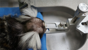
Initially, both the neonatal and adult marmosets had lesions induced in their primary visual cortex. To do so, the marmosets were anaesthetised, administered with antibiotics, and then placed into a stereotaxic frame.
Examples of stereotaxic frames (image sources at bottom of page):


After the marmosets were stabilised, an incision was made following the midline of the skull, and acraniotomy was made, exposing the primary visual cortex of the brain. The dura (the tough outermost membrane enveloping the brain) was then surgically removed, and then the operculumof the primary visual cortex was removed using a blunt scalpel. Sterile absorbable gelatin foam was used to stop bleeding.
Once the flow of blood was stopped, a piece of gelatin film was placed over the surface and the dura was repositioned over the lesion region. Bone fragment was then replaced and affixed with tissue adhesive, and the scalp was sutured. In order to prevent cerebral edema (accumulation of excessive fluid in the brain), the marmosets were given glucose saline, analgesic and dexamethasone (a type of steroid medication).
The marmosets were then recovered in humidicribs before returning to their cages. They were subsequently allowed to ‘recover’ for around 12 months.
Four of the baby marmosets, two of the adult marmosets with lesions induced, and two of the control adult marmosets (with no lesions) were anaesthetised and placed in a stereotaxic frame, and, after receiving an injection directly in the right eyeball, they underwent another craniotomy and had tracer injected into their cortex.
After 14 days, the eight marmosets that received injections were dark adapted for 16 hours, then exposed for 45 minutes of ambient light directly over their cage, before being killed by an overdose of pentobarbitone. They were then transcardially perfused, and cerebral tissues were removed. Brain sections were then examined using immunohistochemistry analysis.
Two of the neonatal lesion marmosets, two adult lesion marmosets, and two control marmosets also had their brains examined through MRI analysis.
All marmosets were killed for the experiment and brain section analysis.
Relevance to Humans
There are major anatomical, genetic, dietetic, environmental, toxic, and immune differences between animals – including marmosets – and humans(1), making them inappropriate for use in studying human brain injury and human disease. Many studies and systematic reviews show that there is discordance between animal and human studies, and that animal ‘models’ fail to mimic clinical disease adequately.(2)(3).
Given the research paper itself identifies previous human trials investigating functional outcomes of damage to the primary visual cortex at different ages, one must seriously question why the researchers engaged in this study did not utilise a human sample to conduct research in this area, and/or use a battery of advanced human biology-based methods of research, in order for results to be directly relevant to human health outcomes.
Funding
The experiment was funded through National Health and Medical Research Council of Australia (NHMRC) project grants (APP1002051 and APP1042893) to researcher J Bourne, as well as by grants from the State Government of Victoria and the Australian Government to the Australian Regenerative Medicine Institute.
What You Can Do
Please use the form below to tell Monash University how disappointed you are with their use of animals in this experiment. You can use the text provided or compose your own (remember polite personalised messages carry more weight).
Your message will be sent via email to the Executive Assistant of the Vice-Chancellor of Monash University.
"*" indicates required fields
Image Sources:
Figure 1 source – Quadrature RF Coil for In Vivo Brain MRI of a Macaque Monkey in a Stereotaxic Head Frame – PubMed (nih.gov)
